
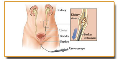
 Ureteroscopy (URS) involves the use of small telescopes that are inserted through the bladder and into the opening of the ureter. The telescope allows visualization of stones in the ureter and/or kidney. This procedure is usually done for treatment of stones stuck in the ureter or in the kidney . Depending on the size of the stone and the diameter of the ureter, the stone may be fragmented and/or removed. The most common method of fragmentation is performed using a laser. The laser is passed through a tiny channel in the telescope and used to fragment the stone into smaller pieces. One benefit of this treatment is the ability to directly visualize the fragmentation process. Moreover, stone fragments can be collected for analysis by passing a stone basket through the telescope and capturing the fragments for removal.
The holmium laser is considered the safest, most effective of all ureteroscopic lithotripsy devices for breaking up stones. It has several advantages. It can transmit its energy through a flexible fibre, which allows its use in both the ureter and the kidney. It is much less likely to cause damage when used within the ureter, and it has the ability to fragment all stones regardless of their composition. The holmium laser also produces significantly smaller fragments compared with other ureteroscopic devices.
Passage of the ureteroscope may result in swelling of the ureter. Therefore, it may be necessary to temporarily leave a small tube inside the ureter called a stent. The stent travels from the kidney, down the ureter and into the bladder. The stent may also assist in the passage of any residual stone fragments. It is not uncommon for some patients to experience some discomfort from the stent. As with ESWL, a minor amount of blood is expected in the urine while the stent is in place. URS is usually performed as an outpatient procedure, requiring use of anesthesia.
URS can be used safely in pregnancy and may be the favored approach in patients with bleeding disorders, on blood thinners or who are morbidly obese. In addition, patients with stones in an abnormally formed kidney may be treated best with URS.
URS is considered safe and effective, but, as with any procedure, risks exist. They include perforation of the ureter and formation of scar tissue that narrows the diameter of the ureter. These adverse events are encountered more frequently when a stone is found lodged or embedded in the wall of the ureter. If perforation occurs, an attempt will be made to place a temporary stent within the ureter to allow the area to heal. In the event that a stent cannot be safely placed, a small tube may need to be placed into the kidney to direct urine away from the damaged area. Fortunately, scar tissue formation in the ureter is rare, occurring in less than 1% of cases. However, when it happens, obstruction to the flow of urine may result. Obstruction can cause pain and may damage the kidney if allowed to persist. Additional intervention is often necessary to treat the narrowed area.
The reasons for a ureteroscopy include the following conditions:
- Frank hematuria
- Abnormal cells found in a urine
- urinary blockage caused by an abnormal narrowing of the ureter
- stone in the ureter
growth, polyp, tumor, or cancer in the ureter